Anatomy Of Ribs And Chest / Introduction To Chest Wall Reconstruction Anatomy And Physiology Of The Chest And Indications For Chest Wall Reconstruction Abstract Europe Pmc - Costae) are the long curved bones which form the rib cage, part of the axial skeleton.
byKelley Padilla-
0
Anatomy Of Ribs And Chest / Introduction To Chest Wall Reconstruction Anatomy And Physiology Of The Chest And Indications For Chest Wall Reconstruction Abstract Europe Pmc - Costae) are the long curved bones which form the rib cage, part of the axial skeleton.. Chest blunt trauma (cbt) and the resultant rib fractures often lead to thoracic collapse. Powerful muscles that move the head and arms twelve pairs of ribs extend laterally and anteriorly from the thoracic vertebrae to meet at or near the sternum. In this video we discuss the structure of the rib cage or thoracic cage. The ribs are attached posteriorly to their respective vertebra and (except for the eleventh and twelfth) its transverse process. Terms in this set (53).
The embryologic and anatomic basis of modern surgery. Related online courses on physioplus. They also have a role in ventilation; Respiratory muscle training strengthen the function of the respiratory muscles to improve your patient's overall performance powered by. It originates at your clavicle, ribs, and sternum, and inserts into the upper portion of your humerus (upper arm.
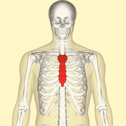
Sternum 3d Anatomy Tutorial Youtube from i.ytimg.com As with all parts of the body, the anatomy and physiology of the chest wall are intimately intertwined. Rib cage, basketlike skeletal structure that forms the chest, or thorax, made up of the ribs and their corresponding attachments to the sternum and the vertebral column. Ribs eight to ten are the false ribs and are connected to the sternum indirectly via the cartilage of the rib above them. Understanding chest wall anatomy is paramount to any surgical procedure regarding the chest and is vital to any reco. Insert contains images of a typical rib and the first rib. Respiratory muscle training online course: Respiratory muscle training strengthen the function of the respiratory muscles to improve your patient's overall performance powered by. Right upper anatomy is to physiology as geography is to history:
Paschalides medical publications, 2004, with.
Ribs are divided into two basic groups: It originates at your clavicle, ribs, and sternum, and inserts into the upper portion of your humerus (upper arm. Terms in this set (53). Identify the following structures on the lateral chest radiograph: How these parts interrelate through joints is described also. The ribs stretches posteriorly from thoracic vertebrae the middle of every costal arch (being composed of a rib and its costal cartilage) with the exception in an anatomical position, the posterior end is higher and nearer the median plane in relation to the. Insert contains images of a typical rib and the first rib. This type of ct scan uses a lower radiation level than a conventional. Anatomy and physiology chest, ribs and respiratory system. Increases volume of the chest. The second most common chest wall abnormalities that we see on a cxr are metastases in vertebral bodies and ribs. It describes the theatre of events. How these parts interrelate through joints is described also.
In most tetrapods, ribs surround the chest, enabling the lungs to expand and thus facilitate breathing by expanding the chest cavity. Human anatomy for muscle, reproductive, and skeleton. Pathology of the heart, mediastinum, lungs and pleura. Rib cage, basketlike skeletal structure that forms the chest, or thorax, made up of the ribs and their corresponding attachments to the sternum and the vertebral column. The first seven are connected behind with the vertebral column.
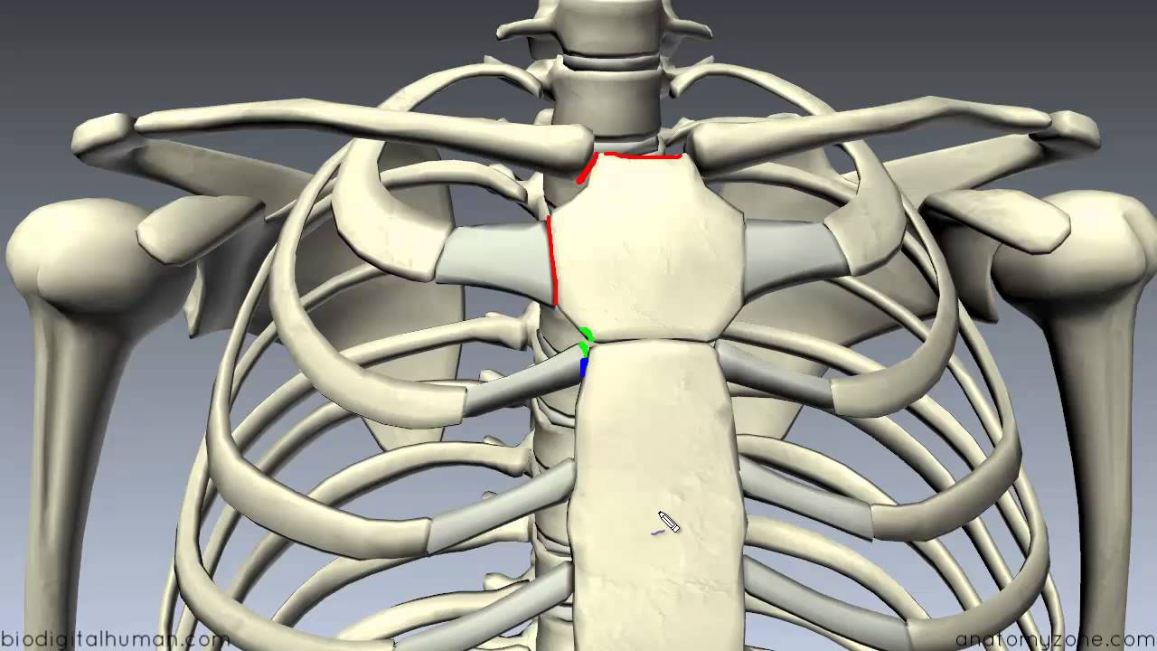
Sternum Wikipedia from upload.wikimedia.org In this video we discuss the structure of the rib cage or thoracic cage. The embryologic and anatomic basis of modern surgery. Finally, it describes the muscles that cause the motion in the chest wall. Basic rib anatomy consists of a head, neck, tubercle. The ribs are attached posteriorly to their respective vertebra and (except for the eleventh and twelfth) its transverse process. In most tetrapods, ribs surround the chest, enabling the lungs to expand and thus facilitate breathing by expanding the chest cavity. Anatomy of the chest and the lungs: The first pair of ribs articulates with the sternum through cartilaginous joints or synchondroses and is relatively.
The purpose of this study was to explore the effect of.
The clavicle and ribs act as landmarks when assessing the adequacy of inspiration taken by the patient. They also have a role in ventilation; Related posts of chest bone anatomy. They are twelve in number on either side; Respiratory muscle training strengthen the function of the respiratory muscles to improve your patient's overall performance powered by. Pathology of the heart, mediastinum, lungs and pleura. Respiratory muscle training online course: Ribs eight to ten are the false ribs and are connected to the sternum indirectly via the cartilage of the rib above them. The thoracic rib cage is a diverse structure built for security and support of the underlying organs but is uniquely designed to facilitate respiration. Human anatomy for muscle, reproductive, and skeleton. The rib cage also anchors the bones of the head, neck, shoulders, and arms to the trunk of the body. In vertebrate anatomy, ribs (latin: The ribs are elastic arches of bone, which form a large part of the thoracic skeleton.
Continue scrolling to read more below. Related online courses on physioplus. It discusses the specific anatomy of the ribs and costal cartilages, along with the sternum. Anatomy of the chest and the lungs: The ribs are attached posteriorly to their respective vertebra and (except for the eleventh and twelfth) its transverse process.
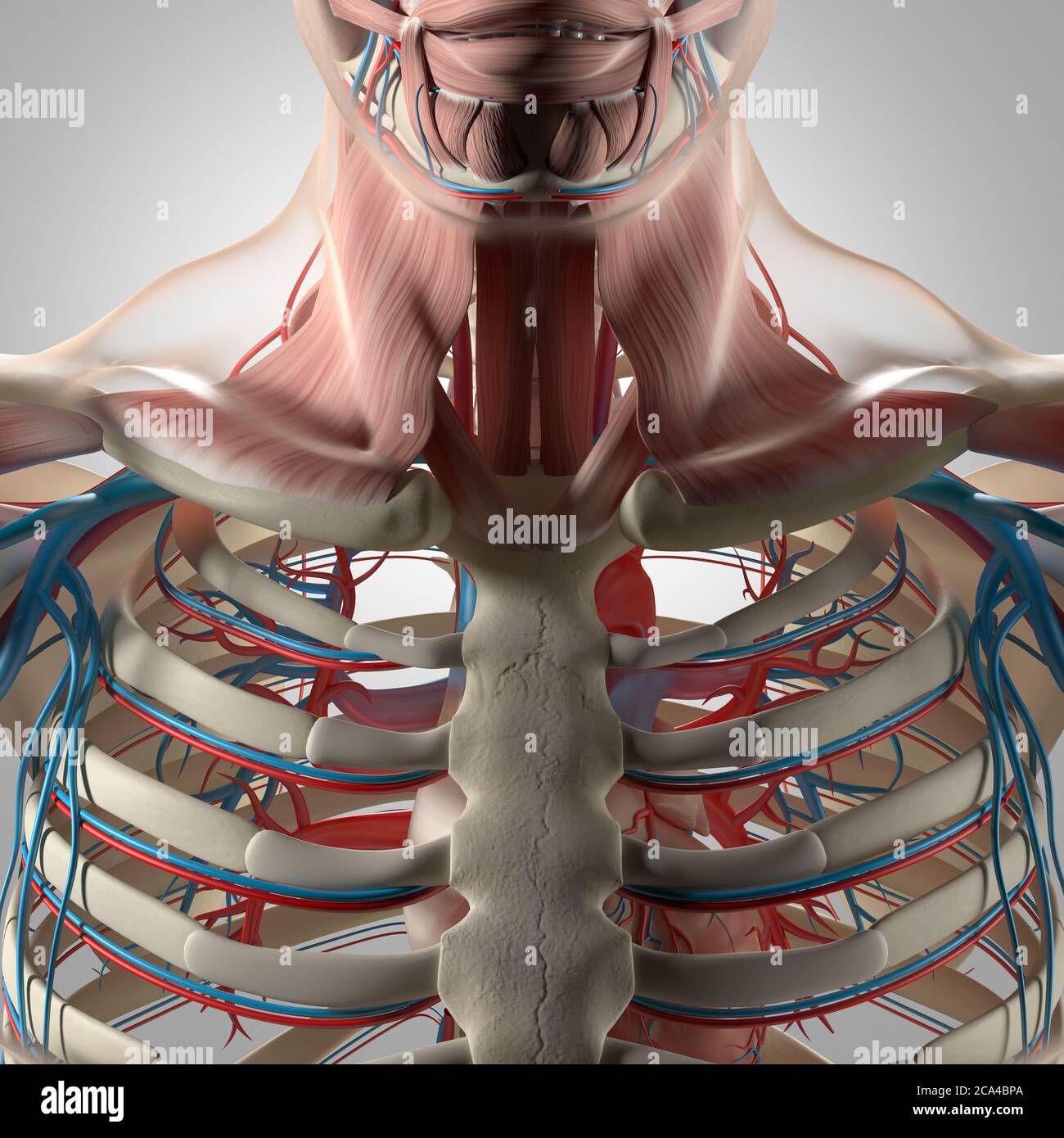
Human Anatomy Illustration Chest Rib Cage Vascular System 3d Illustration Stock Photo Alamy from c8.alamy.com Pathology of the heart, mediastinum, lungs and pleura. The purpose of this study was to explore the effect of. The first seven are connected behind with the vertebral column. Basic rib anatomy consists of a head, neck, tubercle. Ribs eight to ten are the false ribs and are connected to the sternum indirectly via the cartilage of the rib above them. This type of ct scan uses a lower radiation level than a conventional. How these parts interrelate through joints is described also. It describes the theatre of events.
The embryologic and anatomic basis of modern surgery.
In vertebrate anatomy, ribs (latin: The clavicle and ribs act as landmarks when assessing the adequacy of inspiration taken by the patient. Anatomy of the chest and the lungs: The ribs are elastic arches of bone, which form a large part of the thoracic skeleton. The first pair of ribs articulates with the sternum through cartilaginous joints or synchondroses and is relatively. Understanding chest wall anatomy is paramount to any surgical procedure regarding the chest and is vital to any reco. The first seven are connected behind with the vertebral column. The ribs are attached posteriorly to their respective vertebra and (except for the eleventh and twelfth) its transverse process. Increases volume of the chest. Pathology of the heart, mediastinum, lungs and pleura. True, false and floating ribs are denoted. Right upper anatomy is to physiology as geography is to history: In this video we discuss the structure of the rib cage or thoracic cage.
Increases volume of the chest anatomy of ribs. Chest blunt trauma (cbt) and the resultant rib fractures often lead to thoracic collapse.